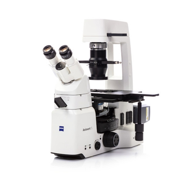
ZEISS Axiovert 5
 ZEISS · Model Axiovert 5
Axiovert 5 delivers excellent results with fast time to image. Simply focus your sample and press a single button to save a crisp image of your cell or tissue culture. For transmitted light and multichannel fluorescence images, this smart microscope automatically adjusts the settings and parameters for you. The overlayed multichannel fluorescence image, including a scale bar, will be saved in the image file metadata. From routine cell culture to research, Axiovert 5 fits seamlessly into your lab and workflow. Various contrasting techniques – DIC, iHMC and phase contrast as well as multichannel fluorescence – open up a huge diversity of applications. Should your work requirements change, Axiovert 5 will adapt, too. Axiovert 5 supports you throughout your daily work. Just keep your eye on your sample: you can access all the main controls with one hand. The Light Manager provides uniform brightness at all magnifications, eliminating manual adjustments to the lamp intensity when you’re changing objectives. To increase the speed and reliability of your cell procedures even more, opt for the AI Cell Confluency and AI Cell Counting modules in Labscope. You’ll get instant, reproducible information on your cells.
Typical Failure Modes
Compiled for this model from its own documentation and established engineering knowledge for this equipment class - not from a verified service record. Confirm against the manufacturer's service manual.
Illumination fault
MediumSymptoms: Lamp / LED out, flickering, uneven or dim field, colour-temperature shift.
Root cause: Halogen bulb or LED module at end of life, a lamp-power-supply fault, or a bad connection.
Focus mechanism fault
MediumSymptoms: Coarse / fine focus slips, drifts, binds, or the stage will not hold height.
Root cause: Worn focus-drive gears or slipping tension clutch.
Stage / condenser movement fault
LowSymptoms: Mechanical stage or condenser will not move smoothly or hold position.
Root cause: Worn stage rack-and-pinion or a loose condenser mount.
Optical contamination / poor image
MediumSymptoms: Haze, spots or debris in the field of view that rotate with an element.
Root cause: Dirty objectives, eyepieces, condenser or camera port.
Camera / imaging fault (if fitted)
LowSymptoms: No image, poor colour, software will not connect.
Root cause: Camera, its cable, or the imaging software.
Serviceable Parts & Consumables
Serviceable items for this model, compiled from its documentation and engineering knowledge for this equipment class. Service class and relative cost are estimates, not manufacturer figures; no part numbers are given. Confirm intervals and pricing against the manufacturer.
Illumination bulb / LED module
Class Frequent wear itemRel. cost $$R&R Fairly easy
Lamp power supply
Class Major service partRel. cost $$R&R Moderate
Focus-drive gear / clutch kit
Class Major service partRel. cost $$R&R Difficult
Stage rack-and-pinion
Class Major service partRel. cost $$R&R Difficult
Objective / eyepiece (as damaged)
Class Major service partRel. cost $$$R&R Fairly easy
| Part | Service class | Relative cost | R&R difficulty |
|---|---|---|---|
| Illumination bulb / LED module | Frequent wear item | $$ | Fairly easy |
| Lamp power supply | Major service part | $$ | Moderate |
| Focus-drive gear / clutch kit | Major service part | $$ | Difficult |
| Stage rack-and-pinion | Major service part | $$ | Difficult |
| Objective / eyepiece (as damaged) | Major service part | $$$ | Fairly easy |
Relative cost: $ lowest · $$$$ highest service-part cost for this equipment class. Not a price.
Troubleshooting Approach
A starting sequence for this model, compiled from its documentation and engineering knowledge for this equipment class. Follow the manufacturer's service documentation for the actual procedure.
Symptom: Dim, uneven or no illumination
- Replace the bulb / LED module (correct type and wattage).
- Check the lamp power supply and connections.
- Re-set Kohler illumination (field and condenser diaphragms, condenser height).
- Clean the collector lens and diffuser.
- Replace the lamp power supply if output is still low.
Symptom: Focus slips or binds
- Adjust the coarse-focus tension clutch per the manual.
- Check for debris or dried grease on the focus ways.
- Inspect the focus-drive gears for wear.
- Re-lubricate or replace the focus-drive kit.
- Confirm the stage holds height under a heavy slide.
Documents & Manuals
No documents available for this device yet.
Related calculators
From BioLab, for work done with this kind of equipment.
Troubleshoot this device with Bio
Describe a symptom or error code. Bio answers from this device's own documented error codes, failure modes, parts, and guides.
Ask Bio