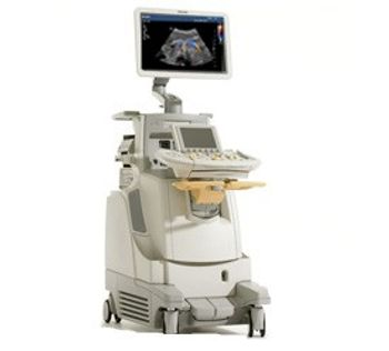
Philips iU22
 Philips Healthcare · Model iU22
Discover the advantages of volume imaging With volume imaging on the iU22 system and the ViewForum workstation, users are now able to view ultrasound in the same format as CT and MR. Virtually re-scan volume data in a familiar workflow, obtain critical information not available with traditional 2D views, take additional measurements, and compare ultrasound studies with other modalities. Reduce failed ultrasound exams by as much as 69% with the C5-1 A recent ultrasound study on technically difficult patients at six sites in North America and Europe using the C5-1 PureWave transducer has shown that this technology can have a very positive impact on an ultrasound lab. Exam times has been reduced up to 38% and studies have shown a reduction in pain and fatigue from scanning in 29% to 85% of exams. Improve early detection and characterization of breast lesions Now with the push of one button, the iU22 system offers tissue aberration correction on breast transducers to compensate for speed of sound variations improving detail resolution and conspicuity of lesion details. Helping users achieve optimum diagnostic performance across the spectrum of breast architecture types - dense to fatty. Reduce ultrasound exam time by up to 50% with SmartExam Introducing the new SmartExam feature, while users perform the ultrasound application, the iU22 remembers every step - all required images for the study, annotation, body markers, mode changes, and quantification requirements. Users can even incorporate 3D data sets into the protocol. Gain a better perspective of needle location during biopsies and ablation The iU22 now has a digital navigation link with the Traxtal PercuNav system for image fusion and improved instrument tracking. The PercuNav system acts like a GPS for medical instruments when guiding soft tissue biopsy and ablation procedures. The combined iU22/PercuNav solution transforms two dimensional patient images into dynamic, fused imaging maps that combine CT or MR imaging with live ultrasound.
Key tips
- ·Never bypass a radiation interlock to "just get the image" - this is one of the few true zero-tolerance rules in BMET work.
- ·Trend image-quality phantom results over time - drift shows up before an outright failure does.
- ·Portable/mobile units take real physical abuse from transport - check wheel locks and cable strain reliefs more often than fixed units.
Typical Failure Modes
Compiled for this model from its own documentation and established engineering knowledge for this equipment class - not from a verified service record. Confirm against the manufacturer's service manual.
Poor 2D image quality across multiple probes
HighSymptoms: Noisy, low-contrast or non-uniform image, missing sector, drop-out that follows the system not the probe.
Root cause: Front-end / beamformer board fault, a probe-port (connector) board fault, or a system-board fault.
Transducer not recognised / probe-port fault
MediumSymptoms: A probe works on one port but not another, or is not detected at all.
Root cause: Worn probe-port connector or a scan-connector board fault.
Will not boot / intermittent shutdown
HighSymptoms: Dead system, hangs at boot, or powers off under load.
Root cause: Power supply / DC converter, the system battery, or a system-board fault.
Control panel / touchscreen or monitor fault
MediumSymptoms: Unresponsive TGC sliders or keys, distorted or blank display.
Root cause: Control-panel board, touchscreen, or the monitor.
Overheating / loud cooling fan
LowSymptoms: Thermal-shutdown warning, fan roar, hot exhaust.
Root cause: Failed or dust-clogged cooling fan or blocked air path.
DICOM / network / storage fault
LowSymptoms: Studies will not send to PACS, images will not archive, worklist empty.
Root cause: Network configuration or a storage-drive fault.
Serviceable Parts & Consumables
Serviceable items for this model, compiled from its documentation and engineering knowledge for this equipment class. Service class and relative cost are estimates, not manufacturer figures; no part numbers are given. Confirm intervals and pricing against the manufacturer.
Front-end / beamformer board
Class Major service partRel. cost $$$$R&R Difficult
Probe-port / scan-connector board
Class Major service partRel. cost $$$R&R Difficult
Power supply / DC converter
Class Major service partRel. cost $$$R&R Difficult
Control-panel / touchscreen assembly
Class Major service partRel. cost $$$R&R Moderate
Cooling fan
Class Major service partRel. cost $R&R Moderate
System hard drive
Class Major service partRel. cost $$R&R Moderate
| Part | Service class | Relative cost | R&R difficulty |
|---|---|---|---|
| Front-end / beamformer board | Major service part | $$$$ | Difficult |
| Probe-port / scan-connector board | Major service part | $$$ | Difficult |
| Power supply / DC converter | Major service part | $$$ | Difficult |
| Control-panel / touchscreen assembly | Major service part | $$$ | Moderate |
| Cooling fan | Major service part | $ | Moderate |
| System hard drive | Major service part | $$ | Moderate |
Relative cost: $ lowest · $$$$ highest service-part cost for this equipment class. Not a price.
PM Checklist
Preventive-maintenance tasks typical of this equipment class. Perform to the manufacturer's procedure and your facility's policy - this is general reference, not a schedule for this specific unit.
- ·Daily QC per modality: image-quality phantom test
- ·Tube and detector inspection
- ·Annual state-required radiation safety survey
- ·Vendor-scheduled preventive maintenance
Troubleshooting Approach
A starting sequence for this model, compiled from its documentation and engineering knowledge for this equipment class. Follow the manufacturer's service documentation for the actual procedure.
Symptom: Poor 2D image quality across multiple probes / channel dropout
- Confirm the fault is present on more than one probe and on a phantom, not just one transducer.
- Inspect the probe-port connectors for bent pins or debris; reseat.
- Run the built-in front-end / channel diagnostics.
- Check cooling - a thermally throttled front end degrades the image.
- Escalate a confirmed beamformer / front-end board fault to the OEM.
Symptom: A transducer is not recognised on one port
- Try that probe on a second port and a known-good probe on the suspect port.
- Clean the port and the probe connector; check for a bent pin.
- Confirm the probe is a supported model for this system software.
- Reboot the system after reseating.
- Replace the probe-port / scan-connector board if the fault stays with the port.
Symptom: Will not boot / intermittent shutdown
- Note any boot / POST beep or on-screen code.
- Check the mains supply and the power-on sequence per the manual.
- Check the power-supply / DC-converter outputs and the system battery.
- Reseat the system boards and drives.
- Refer the power supply or the system board as isolated.
Symptom: Unresponsive control panel / TGC sliders / touchscreen or distorted monitor
- Power-cycle the system.
- Clean around the sliders / trackball; check the control-panel ribbon cable.
- Recalibrate the touchscreen.
- Check the monitor cable and, for a distorted image, the monitor itself.
- Replace the control-panel board or monitor as isolated.
Symptom: Studies will not send to PACS / archive
- Confirm the network cable / Wi-Fi and the DICOM store settings (AE title, IP, port).
- Send a test image and read the DICOM error.
- Check free space on the system drive.
- Verify the PACS accepts from this AE title.
- Restart the DICOM service / system.
Technical Specs
- Cart Based
- 1
- Weight
- 345 lbs
- Clinical Applications
- Colorectal
- Imaging Capabilities
- 3D
- Color-flow Mapping
- 1
- Doppler Modes
- Color, Continuous wave (CW), Pulsed wave (PW)
- Image Display
- 17 in Color Flat Panel
Typical Location
Where this class of equipment is typically deployed - general reference, not this unit's own placement record.
- Radiology / Imaging department
- Emergency Department (portable units)
- Operating Room (C-arms)
- Cath lab
- Bedside ICU (portable X-ray)
Documents & Manuals
Troubleshoot this device with Bio
Describe a symptom or error code. Bio answers from this device's own documented error codes, failure modes, parts, and guides.
Ask Bio