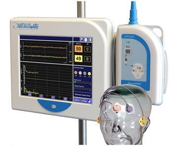
NeuroWave NeuroSENSE NS-901
 NeuroWave · Model NeuroSENSE NS-901
Bilateral Brain Monitoring: The NeuroSENSE Monitor is the first to offer independent bilateral indexes for each brain hemisphere, allowing anesthesia providers to simultaneously monitor brain activity from both sides of the brain during anesthesia or sedation. Instantaneous Response to Patient State Changes: Unlike traditional single-brain monitoring, the NeuroSENSE Monitor responds immediately to changes in patient state, providing real-time insight into anesthetic effects on the brain. Enhanced Clinical Decision-Making: With independent hemispheric measures, clinicians can easily detect discrepancies between brain hemispheres, which could be indicative of underlying pathologies or unrecognized artifacts. Improved Patient Safety: The enhanced bilateral brain activity analysis allows anesthesia providers to make optimal decisions based on a more accurate and reliable view of the patient’s brain state, improving overall patient safety. Real-Time, Simultaneous Monitoring: This system offers instantaneous bilateral brain activity monitoring, enabling doctors in the OR and ICU to quickly identify shifts in sedation or anesthesia levels across both hemispheres for more precise care. Tailored Anesthesia and Sedation: The NeuroSENSE Monitor allows providers to offer personalized sedation or anesthesia protocols based on the real-time data of both hemispheres, optimizing patient outcomes. With its innovative bilateral monitoring capabilities, the NeuroSENSE Monitor enhances clinical decision-making by delivering real-time, comprehensive data for safer and more effective anesthesia and sedation management. This monitor’s advanced features make it an invaluable tool for biomedical engineers and anesthesia providers who require precise and actionable insights into brain activity during surgical procedures or ICU sedation protocols. By incorporating the NeuroSENSE Monitor into patient care, clinicians gain a deeper understanding of the neurophysiological effects of anesthesia, leading to more informed decisions, improved patient safety, and optimized outcomes.
Key tips
- ·Electrode impedance drives data quality more than almost any other single factor - check it before assuming an amplifier fault.
- ·Pre-case verification of intraoperative monitoring equipment isn't optional - a mid-case failure has direct surgical-decision consequences.
Typical Failure Modes
Compiled for this model from its own documentation and established engineering knowledge for this equipment class - not from a verified service record. Confirm against the manufacturer's service manual.
Noisy / high-impedance channels
HighSymptoms: Mains hum, drift or flat channels, persistently high electrode impedance, poor recording quality.
Root cause: Electrode / lead-wire wear, the electrode box (headbox) inputs, or poor skin prep / environment.
Headbox / amplifier fault
MediumSymptoms: A block of channels dead or saturated regardless of electrode, calibration / bio-cal fails.
Root cause: Headbox amplifier or its cable to the acquisition unit.
Amplifier-to-PC link / acquisition fault
MediumSymptoms: Recording drops, amplifier not detected, data gaps.
Root cause: USB / fibre / network link or a driver.
Photic stimulator / video / audio fault (diagnostic EEG/PSG)
LowSymptoms: Photic strobe not firing, no synchronised video, no audio.
Root cause: Photic lamp / trigger, the video camera, or the audio path.
PSG sensor fault (airflow / effort / SpO2 / limb)
LowSymptoms: A PSG channel (thermistor, pressure, RIP belt, SpO2, PLM) flat or noisy.
Root cause: Worn PSG sensor or its cable.
Serviceable Parts & Consumables
Serviceable items for this model, compiled from its documentation and engineering knowledge for this equipment class. Service class and relative cost are estimates, not manufacturer figures; no part numbers are given. Confirm intervals and pricing against the manufacturer.
Electrode lead set / headbox leads
Class Frequent wear itemRel. cost $$R&R Easy
Headbox / amplifier
Class Major service partRel. cost $$$R&R Moderate
Amplifier link cable / interface
Class Periodic replacementRel. cost $$R&R Fairly easy
Photic stimulator lamp (diagnostic)
Class Periodic replacementRel. cost $$R&R Fairly easy
PSG sensor set (airflow / effort / SpO2)
Class Frequent wear itemRel. cost $$R&R Easy
| Part | Service class | Relative cost | R&R difficulty |
|---|---|---|---|
| Electrode lead set / headbox leads | Frequent wear item | $$ | Easy |
| Headbox / amplifier | Major service part | $$$ | Moderate |
| Amplifier link cable / interface | Periodic replacement | $$ | Fairly easy |
| Photic stimulator lamp (diagnostic) | Periodic replacement | $$ | Fairly easy |
| PSG sensor set (airflow / effort / SpO2) | Frequent wear item | $$ | Easy |
Relative cost: $ lowest · $$$$ highest service-part cost for this equipment class. Not a price.
PM Checklist
Preventive-maintenance tasks typical of this equipment class. Perform to the manufacturer's procedure and your facility's policy - this is general reference, not a schedule for this specific unit.
- ·Electrode / lead impedance check
- ·Amplifier calibration
- ·Annual electrical safety test
Troubleshooting Approach
A starting sequence for this model, compiled from its documentation and engineering knowledge for this equipment class. Follow the manufacturer's service documentation for the actual procedure.
Symptom: Noisy or high-impedance channels
- Check and re-prep electrode sites; confirm impedances are balanced and low.
- Swap the affected leads and try different headbox inputs.
- Run the bio-calibration / calibration signal.
- Address mains interference (route cables, check the ground).
- Replace the lead set or headbox as isolated.
Symptom: Amplifier not detected / recording drops
- Reseat the amplifier-to-PC link at both ends.
- Restart the acquisition software and amplifier.
- Try a different port / cable.
- Update the driver / firmware if indicated.
- Replace the link cable or refer the amplifier.
Typical Location
Where this class of equipment is typically deployed - general reference, not this unit's own placement record.
- Neurology / EEG lab
- ICU (continuous EEG monitoring)
- Operating Room (intraoperative monitoring)
Documents & Manuals
Related calculators
From BioLab, for work done with this kind of equipment.
Troubleshoot this device with Bio
Describe a symptom or error code. Bio answers from this device's own documented error codes, failure modes, parts, and guides.
Ask Bio