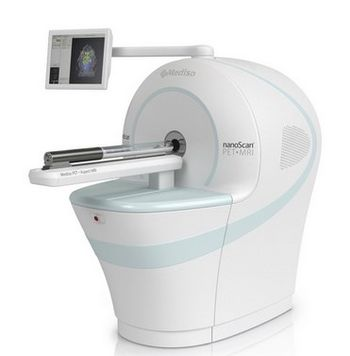
Mediso NanoScan PET/MRI
 Mediso · Model NanoScan PET/MRI
No compromise in imaging performance1: the negligible fringe field from the permanent magnet ensures superior performance of PET subsystem 700 µm spatial resolution with Tera-Tomo 3D PET reconstruction engine with Full Detector Modeling and Attenuation2/Scatter Correction in objects Absolute quantification accuracy over 97% Versatile soft tissue contrast for the best Volume of Interest definitions Determine the volume of tumors: for characterizing the size, vascularization and heterogeneity Detailed structural information in the brain reveals exact distribution of neurological PET tracers Reduced radiation exposure of the animals Anatomical information obtained with non-ionizing radiation (especially important for treatment models) Compact and easy-to-use design does not require special training/education There is no need for long calibrations after start-up There is no waiting time between PET and MRI acquisitions Quick and easy installation Superior PET detector characteristics: Physical performance matches animal imaging performance High-density, ultra-small detectors in tight packing with finest LYSO detector elements in the industry with smallest pitch Increased crystal thickness for highest sensitivity Measure even the finest details Tera-Tomo 3D PET reconstruction engine brings PET resolution close to the physical limits with outstanding quantification and better signal recovery and signal-to-noise ratio Features Monte Carlo system matrix modeling and Depth of Interaction correction; incorporates attenuation1, scatter, positron range, random and dead time corrections Reconstruction running on multiple GPUs (Graphical Processing Units) delivering shorter computational time Perform Acquisition and Reconstruction simultaneously with the powerful workstations including DICOM server and multi-modality display and analysis suite Highest ever resolution in PET State-of-the-art Tera-Tomo 3D PET image reconstruction engine Extremely fast workflow of data handling and image processing Plug-and-publish: totally shielded, cryogen-free and maintenance-free MRI, no room modifications or shielding needed, just a standard public voltage plug Silent operation, ideal for awake animal brain studies Easy installation, compact size: The whole system including PDU and spectrometer rack fits in any biological lab and needs 2 less than 3 m of lab floor space One-click MultiCell animal anesthesia / imaging bed support system.
Key tips
- ·Never bypass a radiation interlock to "just get the image" - this is one of the few true zero-tolerance rules in BMET work.
- ·Trend image-quality phantom results over time - drift shows up before an outright failure does.
- ·Portable/mobile units take real physical abuse from transport - check wheel locks and cable strain reliefs more often than fixed units.
Typical Failure Modes
Compiled for this model from its own documentation and established engineering knowledge for this equipment class - not from a verified service record. Confirm against the manufacturer's service manual.
RF coil fault / poor SNR
HighSymptoms: Low signal-to-noise, shading, or a dead region in images; coil not detected or fails the coil check.
Root cause: Damaged RF coil element, a coil-cable / connector fault, or a preamp fault.
Gradient fault / image distortion
HighSymptoms: Geometric distortion, gradient-amplifier faults, acoustic change, over-temperature trips.
Root cause: Gradient-amplifier fault, gradient-coil cooling problem, or a gradient-coil fault.
Cryogen / cold-head / compressor fault
HighSymptoms: Rising helium boil-off, cold-head noise or stall, He level dropping, compressor alarms.
Root cause: Cold-head at end of life, a helium-compressor fault, or a chilled-water problem.
Chilled water / cooling fault
MediumSymptoms: System-cooling alarms, gradient or RF over-temperature, scans aborting.
Root cause: Facility chilled-water supply, the heat exchanger, or internal pumps.
Patient table / positioning fault
MediumSymptoms: Table will not dock, drive or index; landmarking errors.
Root cause: Table drive, docking sensor, or encoder fault.
Reconstruction / host / network fault
MediumSymptoms: Recon errors, host hangs, images will not reach PACS.
Root cause: Recon or host computer fault, or the network / storage.
Serviceable Parts & Consumables
Serviceable items for this model, compiled from its documentation and engineering knowledge for this equipment class. Service class and relative cost are estimates, not manufacturer figures; no part numbers are given. Confirm intervals and pricing against the manufacturer.
RF receive coil (exchange)
Class Major service partRel. cost $$$$R&R Moderate
Cold head
Planned replacement; He boil-off rises as it wears.
Class Periodic replacementRel. cost $$$$R&R Difficult
Helium compressor / adsorber
Class Major service partRel. cost $$$$R&R Difficult
Gradient amplifier module
Class Major service partRel. cost $$$$R&R Expert-level
Chilled-water pump / heat exchanger
Class Major service partRel. cost $$$R&R Difficult
Patient table drive / encoder
Class Major service partRel. cost $$$R&R Difficult
| Part | Service class | Relative cost | R&R difficulty |
|---|---|---|---|
| RF receive coil (exchange) | Major service part | $$$$ | Moderate |
| Cold head Planned replacement; He boil-off rises as it wears. | Periodic replacement | $$$$ | Difficult |
| Helium compressor / adsorber | Major service part | $$$$ | Difficult |
| Gradient amplifier module | Major service part | $$$$ | Expert-level |
| Chilled-water pump / heat exchanger | Major service part | $$$ | Difficult |
| Patient table drive / encoder | Major service part | $$$ | Difficult |
Relative cost: $ lowest · $$$$ highest service-part cost for this equipment class. Not a price.
PM Checklist
Preventive-maintenance tasks typical of this equipment class. Perform to the manufacturer's procedure and your facility's policy - this is general reference, not a schedule for this specific unit.
- ·Daily QC per modality: image-quality phantom test
- ·Tube and detector inspection
- ·Annual state-required radiation safety survey
- ·Vendor-scheduled preventive maintenance
Troubleshooting Approach
A starting sequence for this model, compiled from its documentation and engineering knowledge for this equipment class. Follow the manufacturer's service documentation for the actual procedure.
Symptom: Poor SNR or a dead region with one coil
- Swap in a known-good coil of the same type and repeat the scan.
- Run the coil-check / coil-QA tool.
- Inspect the coil housing and cable for damage; reseat the connector.
- Scan the ACR / QA phantom and compare SNR to baseline.
- Quarantine and exchange the faulty coil.
Symptom: Rising helium boil-off or cold-head noise
- Log the helium level and boil-off trend daily.
- Check the cold-head sound and the compressor pressures against spec.
- Confirm the chilled-water supply temperature and flow.
- Schedule a cold-head / compressor service before the level reaches the OEM action point.
- Never let the magnet approach a quench threshold - escalate urgently.
Typical Location
Where this class of equipment is typically deployed - general reference, not this unit's own placement record.
- Radiology / Imaging department
- Emergency Department (portable units)
- Operating Room (C-arms)
- Cath lab
- Bedside ICU (portable X-ray)
Documents & Manuals
Troubleshoot this device with Bio
Describe a symptom or error code. Bio answers from this device's own documented error codes, failure modes, parts, and guides.
Ask Bio