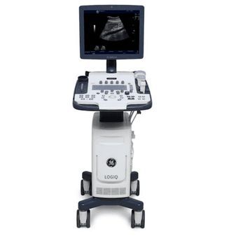
GE HealthCare Logiq V5
 GE HealthCare · Model Logiq V5
Advanced Imaging & Clinical Evaluation It incorporates a number of technologies for providing clear, high-quality images. These include Real Time Speckle Reduction Imaging (SRI-HD) to reduce noise and MLA4 technology to increase frame rate. When the MLA4 technology is used in combination with Color Doppler, it facilitates study of moving organs and blood flow data. In addition, the Logiq V5 uses the CrossXBeam imaging technique for enhancing tissue interfaces and border differentiation with compound imaging technology. The Logiq V5 ultrasound's advanced clinical evaluation capabilities are augmented by Raw Data Imaging (TruScan architecture) for image optimization, Tissue Velocity Imaging (TVI) for assessing heart function with myocardial Doppler imaging, and Easy 3D technology for visualizing volumetric data sets. The V5 Expert also provides other features like Coded Harmonics, Tissue Harmonics, Pulse Wave, and Power Doppler. Simplified Workflow A combination of several features allows for significant efficiencies and streamlines workflow. Customizable menu, user defined keys for workflow customization, 'Switch Probe' button for a quick switch, built-in imaging, and measurement presets for hassle-free scanning are just some workflow optimization techniques. Automation The GE Logiq V5 promotes further efficiencies with auto optimization features like SonoBiometry for fast completion of key fetal measurements, Auto Intima-Media Thickness (IMT) for automatic edge detection, and TruScan Architecture (GE Raw Data) for flexibility in readjusting parameters and re-measuring images. Efficient Ergonomics The Logiq V5 Expert's compact, slim design makes it ideal for clinics and other constricted spaces. Moreover, being lightweight, the V5 is easy to transport. It has a large 15" LCD screen with a 4-way tilt. The GE Logiq V5 is a good choice if you're looking for an ideal balance of clinical capability and affordability.
Key tips
- ·Never bypass a radiation interlock to "just get the image" - this is one of the few true zero-tolerance rules in BMET work.
- ·Trend image-quality phantom results over time - drift shows up before an outright failure does.
- ·Portable/mobile units take real physical abuse from transport - check wheel locks and cable strain reliefs more often than fixed units.
Typical Failure Modes
Compiled for this model from its own documentation and established engineering knowledge for this equipment class - not from a verified service record. Confirm against the manufacturer's service manual.
Will not power on or unexpected shutdown
HighSymptoms: The medical imaging is dead, reboots, or shuts down mid-use.
Root cause: Mains lead / fuse, an aged battery, or a power-supply board fault.
Battery will not hold charge
MediumSymptoms: Short run time on battery; low-battery alarm soon after a full charge.
Root cause: Aged rechargeable battery pack or a charger fault.
Display / control-panel fault
MediumSymptoms: Blank, dim or distorted display; unresponsive keys or touchscreen.
Root cause: Display, backlight, or control-board fault.
Fails self-test or calibration
MediumSymptoms: Power-on self-test or the scheduled performance check fails or is out of tolerance.
Root cause: Sensor drift or an internal board fault; recalibration or repair needed.
Serviceable Parts & Consumables
Serviceable items for this model, compiled from its documentation and engineering knowledge for this equipment class. Service class and relative cost are estimates, not manufacturer figures; no part numbers are given. Confirm intervals and pricing against the manufacturer.
Rechargeable battery pack
Class Periodic replacementRel. cost $$R&R Fairly easy
Power supply / mains board
Class Major service partRel. cost $$$R&R Difficult
Display / control-panel assembly
Class Major service partRel. cost $$$R&R Moderate
Cooling fan
Class Major service partRel. cost $R&R Moderate
| Part | Service class | Relative cost | R&R difficulty |
|---|---|---|---|
| Rechargeable battery pack | Periodic replacement | $$ | Fairly easy |
| Power supply / mains board | Major service part | $$$ | Difficult |
| Display / control-panel assembly | Major service part | $$$ | Moderate |
| Cooling fan | Major service part | $ | Moderate |
Relative cost: $ lowest · $$$$ highest service-part cost for this equipment class. Not a price.
PM Checklist
Preventive-maintenance tasks typical of this equipment class. Perform to the manufacturer's procedure and your facility's policy - this is general reference, not a schedule for this specific unit.
- ·Daily QC per modality: image-quality phantom test
- ·Tube and detector inspection
- ·Annual state-required radiation safety survey
- ·Vendor-scheduled preventive maintenance
Troubleshooting Approach
A starting sequence for this model, compiled from its documentation and engineering knowledge for this equipment class. Follow the manufacturer's service documentation for the actual procedure.
Symptom: Will not power on
- Confirm the outlet, mains lead and fuse.
- Try on battery, then on mains only, to isolate the supply.
- Look for a power indicator or POST code.
- Replace the fuse / battery / power supply as isolated.
- Run the self-test after any repair.
Symptom: Fails self-test or calibration
- Note the exact failure message / code.
- Power-cycle and repeat the self-test.
- Verify against a reference / simulator where one applies.
- Recalibrate per the service manual.
- Refer the affected board for service if it will not hold calibration.
Typical Location
Where this class of equipment is typically deployed - general reference, not this unit's own placement record.
- Radiology / Imaging department
- Emergency Department (portable units)
- Operating Room (C-arms)
- Cath lab
- Bedside ICU (portable X-ray)
Documents & Manuals
No documents available for this device yet.
Troubleshoot this device with Bio
Describe a symptom or error code. Bio answers from this device's own documented error codes, failure modes, parts, and guides.
Ask Bio