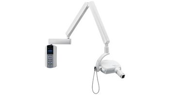
Acteon Group X-Mind Unity
 Acteon Group · Model X-Mind Unity
1. A sharp and contrasted image The x-mind unity has a.4 focal spot. It has several configurable radiological settings notably: The anodic voltage (60,65 and 70 kV) The anodic current (from 4 to 7 mA) These parameters ensure a sharp and contrasted image 2. Up to 52% less radiation with ACE. This technology applied to X-Mind Unity allows the Sopix Inside sensor to start and stop the generator, thus avoiding all risk of over exposing the image and patient as well as unnecessary re-takes of acquisitions. The patient only received the necessary dose, adapted to his dental morphology. 3. Ergonomic and simplistic use, designed to your image X-Mind Unity is pre-wired to allow perfect integration of the Sopix sensor in the future. Sopix inside enables integration without any visible cables. Sopix inside connects effortlessly to the X-Mind Unity without technical assistance within a minute. Exceptional working comfort due to its ergonomically designed holde, there is no danger of the sensor falling or breaking, plus the sensor always remains within hands. 4. Quiet mind for all with exclusive traceability The dose received by the patient after each exposure appears on the timer's screen. When using Sopix inside with X-Mind unity, AIS software systematically records the X-Mind Unity settings as well las the effective dose received by the patient for each acquisition This ensures permanent traceability for every patient. 5. Advanced functionality for intuitive navigation The Acteon Imaging software offers intuitive navigation and advanced functionality. It alone lets you manage all of your images, from scanning to viewing images from all Acteon Imaging devices (CBCT, Panoramic, intraoral digital x-ray system, intraoral camera, etc.) and much more.
Key tips
- ·Biological-indicator verification applies to dental sterilizers exactly the same as hospital CSSD - no exception for smaller units.
- ·"Sounds different" is often the first real report of handpiece bearing wear, not an outright failure - investigate early rather than waiting.
Typical Failure Modes
Compiled for this model from its own documentation and established engineering knowledge for this equipment class - not from a verified service record. Confirm against the manufacturer's service manual.
Output / performance below spec
HighSymptoms: Compressor will not build pressure / cycles constantly; vacuum weak; curing light low irradiance; scanner poor scans.
Root cause: Wear in the core mechanism (compressor head, vacuum pump, LED / lamp, scanner optics) or a control fault.
Water / moisture / drain fault (compressor)
MediumSymptoms: Water in the air lines, drain not working, desiccant exhausted.
Root cause: Auto-drain fault or an exhausted air-dryer / desiccant.
Overheating / thermal cut-out
MediumSymptoms: Unit stops after a period, hot, restarts when cool.
Root cause: Cooling-fan fault, a blocked intake, or a worn motor.
Control / switch / timer fault
MediumSymptoms: Will not start / stop on demand, timer or mode selection unresponsive.
Root cause: Pressure switch, timer, or control board.
Noise / vibration
LowSymptoms: Loud operation, rattling, walking across the floor.
Root cause: Worn mounts, bearings, or loose panels.
Serviceable Parts & Consumables
Serviceable items for this model, compiled from its documentation and engineering knowledge for this equipment class. Service class and relative cost are estimates, not manufacturer figures; no part numbers are given. Confirm intervals and pricing against the manufacturer.
Core mechanism rebuild kit (compressor head / pump / LED)
Class Periodic replacementRel. cost $$R&R Moderate
Air-dryer / desiccant / auto-drain (compressor)
Class Frequent wear itemRel. cost $R&R Fairly easy
Cooling fan
Class Periodic replacementRel. cost $R&R Fairly easy
Pressure switch / timer / control board
Class Major service partRel. cost $$R&R Moderate
Anti-vibration mounts / bearings
Class Periodic replacementRel. cost $R&R Moderate
| Part | Service class | Relative cost | R&R difficulty |
|---|---|---|---|
| Core mechanism rebuild kit (compressor head / pump / LED) | Periodic replacement | $$ | Moderate |
| Air-dryer / desiccant / auto-drain (compressor) | Frequent wear item | $ | Fairly easy |
| Cooling fan | Periodic replacement | $ | Fairly easy |
| Pressure switch / timer / control board | Major service part | $$ | Moderate |
| Anti-vibration mounts / bearings | Periodic replacement | $ | Moderate |
Relative cost: $ lowest · $$$$ highest service-part cost for this equipment class. Not a price.
PM Checklist
Preventive-maintenance tasks typical of this equipment class. Perform to the manufacturer's procedure and your facility's policy - this is general reference, not a schedule for this specific unit.
- ·Daily handpiece lubrication and autoclave cycle verification
- ·Compressor moisture-trap draining
- ·Annual full PM
Error Codes
Extracted directly from this unit's own service manual.
E101memory fail EEPROM fault NO
E102memory fail I2C bus EEPROM fault NO
E103memory fail EEPROM DMA readings fault NO
E104memory fail EEPROM DMA writings fault NO
E105no response from generator Lost communication between inverter and generator NO
E106wrong response from generator NAK response from inverter YES
E107emission start fail YES
E108emission too long YES
E109button released beforehand YES
E110wrong use of button Exposure button pressed when “ready” mode not active.
E111keyboard pressed at boot Keyboard pressed during boot time NO
E112button pushed at boot Exposure button pressed beforehand NO
E113wrong use of keyboard Keys pressed during an exposure YES
E114button release timeout Exposure button released beforehand after an exposure YES
E115wrong parameters in generator Exposure parameters (kV, mA, exposure time, mode) does not match with the ones set in the control box YES
E116generator reset during emission inverter has been resetted during an exposure YES
E117calibration start fail Calibration YES
E118calibration too long Calibration time too long YES
E119button pushed during cooling Exposure button pressed beforehand pushed during a functionality exposure YES
E120cooling time disabled Functionality pause disabled YES
E121button pushed during stand-by Exposure button pressed beforehand during stand-by YES
E122memory fail Parameters out of range NO
E123power relay fail power relay failure (relay OFF) NO
E124power relay fail power relay failure (relay ON) NO
E125power board fail power board failure NO
E126power board fail power board failure NO
E127power board fail power board failure NO
E128power board fail power board failure NO
E129wireless unit not connected wireless receiver board not connected YES
E132system error hardware error NO
E201generator memory Inverter EEPROM error NO
E202generator memory Inverter I2C EEPROM error NO Code Message Description Peut être réinitialisé
E203generator memory Inverter EEPROM DMA readings fault NO
E204generator memory Inverter EEPROM DMA writings fault NO
E205generator not calibrated Calibration not yet done YES
E206tube voltage too low Anodic voltage too low YES
E207tube voltage too high Anodic voltage too high YES
E208electrical discharge on hv YES
E209tube current too low Anodic current too low YES
E210tube current too high Anodic current too high YES
E211filament voltage too low Filament voltage too low YES
E212filament voltage too high Filament voltage too high YES
E213tube unit not connected High voltage unit not connected NO
E214button released beforehand Enable signal released beforehand during an exposure YES
E215generator internal power supply 12V internal voltage too low NO
E216generator internal power supply 12V internal voltage too high NO
E217generator mode Wrong exposure enabling signal activation YES
E218safe circuit fail Security interlock fault NO
E219tube current too low Mean anodic current too low YES
E220tube current too high Mean anodic current too high YES
E221no tube voltage feedback Missing HV feedback NO
E222ACE acquisition fail X-rays not detected by Sopix YES
E223tube unit temperature too high Monoblock over temperature YES
E224tube unit temperature too low Monoblock under temperature YES
E225tube unit temp. sensor fail Monoblock temperature sensor fault (open circuit) YES
E226tube unit temp. sensor fail Monoblock temperature sensor fault (short circuit) YES
E227voltage reference fail generator voltage reference too low YES
E228voltage reference fail generator voltage reference too high YES
E232system error hardware error NO For the errors that can’t be cleared by the reset button on the keypad, please contact your installer or your local Acteon customer service
Troubleshooting Approach
A starting sequence for this model, compiled from its documentation and engineering knowledge for this equipment class. Follow the manufacturer's service documentation for the actual procedure.
Symptom: Detector not connecting / images not received / link drops
- Check the detector battery charge (wireless) or reseat the tether.
- Confirm the Wi-Fi access point, channel and range.
- Reboot the detector and the acquisition workstation.
- Try a known-good detector if the site has one.
- Escalate a confirmed detector-electronics fault to the OEM.
Symptom: Fixed-pattern artefacts / non-uniformity / dead pixels or lines / failed calibration
- Run the detector gain / offset calibration and the dead-pixel map.
- Inspect the detector for drop / crush damage.
- Expose a uniform phantom and check uniformity.
- Confirm the generator and AEC are within spec with an X-ray analyser.
- Exchange the detector if calibration will not hold.
Symptom: No or inconsistent exposure / generator error / kV-mAs inaccuracy
- Note the exact generator error / fault code.
- Confirm the mains supply is within the generator's spec (line-voltage compensation).
- Check the HT cables and the exposure contactor.
- Confirm the tube is not in a thermal lockout.
- Escalate a confirmed inverter / HV-tank / tube / filament fault; verify output with an analyser after any repair.
Symptom: AEC (phototimer) images consistently over- or under-exposed
- Verify the AEC response with a phantom at two thicknesses.
- Check the AEC chamber connections and the selected chambers / density.
- Recalibrate the AEC per the service procedure.
- Compare against a manual technique.
- Replace the AEC chamber if it will not calibrate.
Symptom: Tube stand / table / wall bucky will not track, detent or auto-position
- Check the drive, brake, detents and counterbalance for that axis.
- Inspect the position encoder / potentiometer.
- Check the auto-positioning interface and cabling.
- Adjust or replace the brake / drive hardware.
- Confirm smooth motion and reliable detents / SID.
Typical Location
Where this class of equipment is typically deployed - general reference, not this unit's own placement record.
- Dental operatory
- Oral surgery suite
- CSSD (dental sterilizers)
Documents & Manuals
Troubleshoot this device with Bio
Describe a symptom or error code. Bio answers from this device's own documented error codes, failure modes, parts, and guides.
Ask Bio